Endometrial Cancer
Introduction
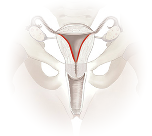
Endometrial cancer, also known as uterine cancer, is the abnormal growth of any cells that originate from the endometrium, the inner lining of the uterus (womb). These abnormal cells grow uncontrollably, forming a mass called a tumour. Endometrial cancer is the fourth most common cancer diagnosed in females in Singapore.*
Certain tumours of the uterus can however be benign, such as a fibroid, a polyp, or endometriosis. These are usually not life-threatening and can be treated or removed. They do not invade neighbouring tissues or spread to other parts of the body.
Malignant growths, which are cancerous, can invade nearby tissues and organs such as the vagina and spread to other parts of the body. The cancer cells break out of the uterine tumour and travel via lymph vessels to nearby lymph nodes. They can also spread through the blood vessels to other organs such as the lung, liver, or brain, forming tumours in these new locations.
* Singapore Cancer Registry Annual Registry report, Trends in Cancer Incidence in Singapore 2023
Risk Factors
- Age (the risk of uterine cancer increases as a woman gets older)
- Obesity (fatty tissue produces additional estrogen)
- Diabetes (women with diabetes may have as much as 4 times the risk of uterine cancer)
- Women who have never been pregnant
- Women who had their first menstrual period before the age of 12 or had undergone menopause after the age of 55
- Women with irregular/infrequent menses i.e. less than 4 menses per year, which is indicative of a failure to ovulate
- Women who have a personal history of breast or ovarian cancer
- Women who have family members with uterine cancer or a family history of Hereditary Non-Polyposis Colorectal Cancer (HNPCC) or Lynch Syndrome
- Female breast cancer patients who have been treated with Tamoxifen
- Women with polycystic ovarian syndrome
- Women who have used an intrauterine device
Signs & Symptoms
Women with uterine cancer tend to display symptoms at an early stage, such as:
- Unusual vaginal bleeding (e.g., post-menopausal bleeding, irregular menstrual bleeding, abnormally heavy bleeding/Menorrhagia)
- Vaginal spotting
- Pink or white, watery vaginal discharge
- Pain during sexual intercourse
- Pain during urination
- Occasional pelvic pain
Early Detection and Screening
Screening for uterine cancer is not recommended for women with an average or increased risk for uterine cancer. Women with high risk (with or at risk of hereditary non-polyposis colorectal cancer) should be offered annual screening for uterine cancer with transvaginal ultrasound and endometrial biopsy by age 30-35.
Diagnosis
Pelvic Examination
Performed by a doctor on the uterus, vagina, ovaries, and rectum to look for possible lumps or any unusual findings.
Ultrasound Scan
Using sound waves to create an image of the pelvic region to identify abnormalities in the endometrium or any abnormally-sized/shaped organs. A trans-vaginal ultrasound may be performed to get a better view of the uterus by inserting the ultrasound device into the vagina.
Endometrial Biopsy
This is required for a definite diagnosis. A small amount of uterine tissue is removed using suction from a thin tube inserted through the cervix into the uterus. This tissue is examined under the microscope by a pathologist to look for cancerous cells.
Dilatation and Curettage (D&C)
This procedure removes uterine tissue with the aid of hysteroscopy, allowing the doctor to visualise the uterine lining using a thin lighted tube inserted through the cervix. Additional testing may be required to determine the progress of cancer and to allow for the proper categorisation of the cancer by stage before an appropriate type of treatment is chosen.
Treatment
Surgery
Surgery is the most common treatment for uterine cancer. The surgeon removes the uterus (hysterectomy), cervix, and possibly other nearby tissue such as the ovaries, fallopian tubes, and local lymph nodes depending on the spread of the cancer.
Radiation Therapy
Radiation therapy is an option for all stages of uterine cancer and may be utilised before or after surgery. This method utilises high-energy rays to destroy cancer cells in the treated area.
Chemotherapy
Chemotherapy utilises drugs to destroy cancer cells and is usually given after surgery. The drugs are delivered either intravenously or swallowed in the form of a pill or capsule. A combination of different drugs may sometimes be administered at the same time for optimal treatment.
Hormone Therapy
Hormone therapy is used to slow the growth of certain types of uterine cancer. These are generally adenocarcinomas and are grade I or II tumours. This method involves delivering the sex hormone progesterone through a pill.